+
Transparent Wall - Individual Blister Pack
Ideal for fluorescence microscopy, confocal (laser) microscopy, high-resolution image analysis, cell culture, and electrophysiology.
Transparent Wall - Individual Blister Pack
Ideal for fluorescence microscopy, confocal (laser) microscopy, high-resolution image analysis, cell culture, and electrophysiology.
This series of sterile, optical quality, glass bottom dishes provide exceptional imaging quality for many applications requiring the use of inverted microscopes. Conventional plastic or glass petri dishes and chambers limit the use of the inverted microscopy for many applications because they require a long working distance objective available only in lower magnifications. Moreover, plastic dishes cannot be used for DIC or any polarization microscopes due to their inherent birefringence. These dishes eliminate these problems, making them ideal for applications such as: patch clamp recordings where fluorescent-tagged (GFP) receptors can be identified for selective study and many other fluorescent microscopy applications which require low background scattering of light and reduce intrinsic "auto" fluorescence that occurs when using standard plastic dishes. Furthermore, the glass bottom is designed flush with the whole base of the dish, allowing direct contact between the entire glass surface and the warming stage. This eliminates any air gaps that may otherwise exist, optimizing heat-transfer, and allowing hojmogenous heating and cooling of the glass bottom.
Features
Non-cytotoxic, non-pyrogenic, gamma irradiated (EN 552) and sterile until opened.
Production is carried out in a Class 10,000 clean room, certified under EN46001, EN46002, ISO9001, and ISO 9002.
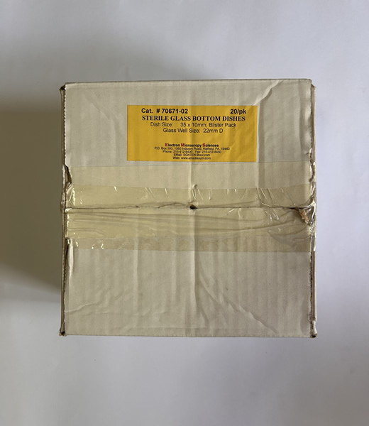
Dish diameters: 35 and 50 mm, with internal glass well diameter of 12, 22, 33, or 40 mm.
Available individually packed in a medical-style blister or in sleeves of 20 dishes.
Optical Properties
Wide optical range of transmission- UV, IV, and IR (300nm to 2500 nm).
Flat (0.17 mm thick) optical quality glass bottom provides the following advantages:
Use of a much shorter working distance immersion objective
Larger numerical aperture (NA) objectives
Higher magnification (up to 100x)
Applications
Confocal (laser) microscopy.
Two-/multi photon confocal microscopy.
DIC and polarization microscopy.
High-resolution imaging system.
Infrared imaging (e.g., brain slide recordings).
Scientific Fields
Cell Biology: STEM cell research, cell growth cycle monitoring, cell culture.
Protein Chemistry: Green fluorescent protein identification of histotags, etc.
Molecular Biology: Phenotyping and other complex genetic research.
Neurology: Electrolyte studies, axon growth.
Pathology: Particle and cell morphology.
Fertility/Infertility: Biopsies, ICSI, spermatid identification.
2 İş Gününde Kargo
Güvenli Alışveriş
%100 Memnuniyet
7/24 Destek